Key takeaways
Dental imaging — panoramic (OPG) X-rays, cephalometric X-rays and CBCT scans — lets your dentist assess teeth, jawbone and nerve positions before treatment, so implant placement, wisdom tooth removal, root canal treatment and orthognathic surgery can be planned on solid evidence. Digital 3D intraoral scanning replaces part of the traditional impression process with an optical scan that feeds directly into crown, bridge and clear-aligner design. Each type of imaging has its own indications and radiation considerations; your dentist recommends imaging only when it is clinically needed.
Patients often ask two opposite questions: "Isn't a simple X-ray enough — why a CT scan?" and "Is all this really necessary?" This page explains what each type of dental imaging shows and when it is actually needed, so that when your dentist recommends a scan, you understand the clinical reasoning and can make an informed decision.
Panoramic radiograph (OPG): the whole mouth in one image
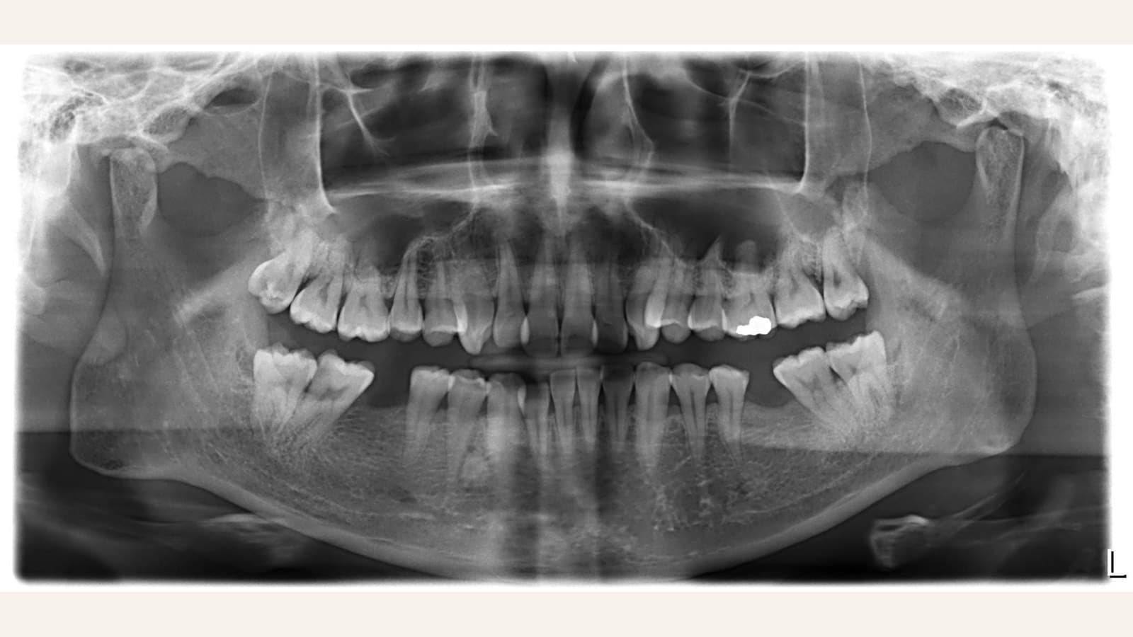
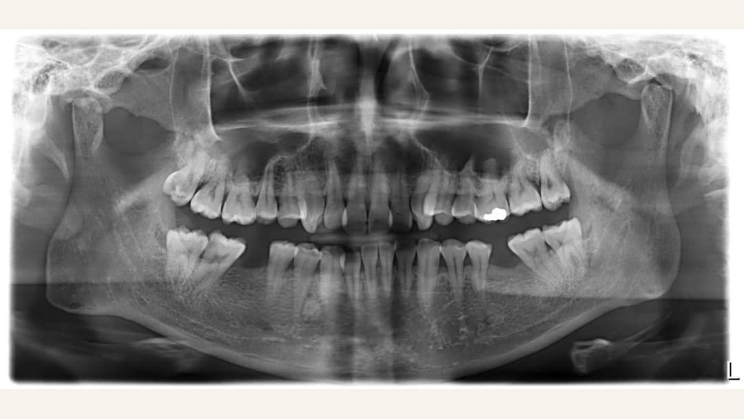
The OPG is one of the most common dental images. The machine rotates around your head and captures the upper and lower jaws, all teeth, the jawbone and the jaw joints in a single flat image.
Typical uses include: full-mouth assessment at a first visit, evaluating wisdom tooth position and angulation, reviewing overall periodontal bone levels, and screening for impacted teeth or jawbone shadows. Compared with taking many small films, an OPG gives a whole-mouth overview in one exposure, at a low radiation dose.
Cephalometric X-ray: the foundation of orthodontic and jaw-surgery planning
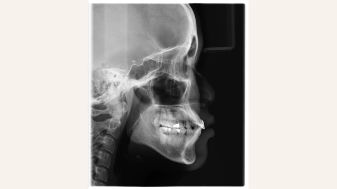
A cephalometric X-ray captures the whole skull from the side (or front), showing the relative positions of the teeth, the upper and lower jaws and the facial soft tissues. Orthodontists measure a series of angles and proportions on this image to distinguish dental from skeletal problems, plan braces or aligner treatment or orthognathic surgery, and track progress during treatment.
Put simply: whether orthodontic treatment is actually progressing is judged by objective measurements on cephalometric films — not by eye.
CBCT: three-dimensional imaging
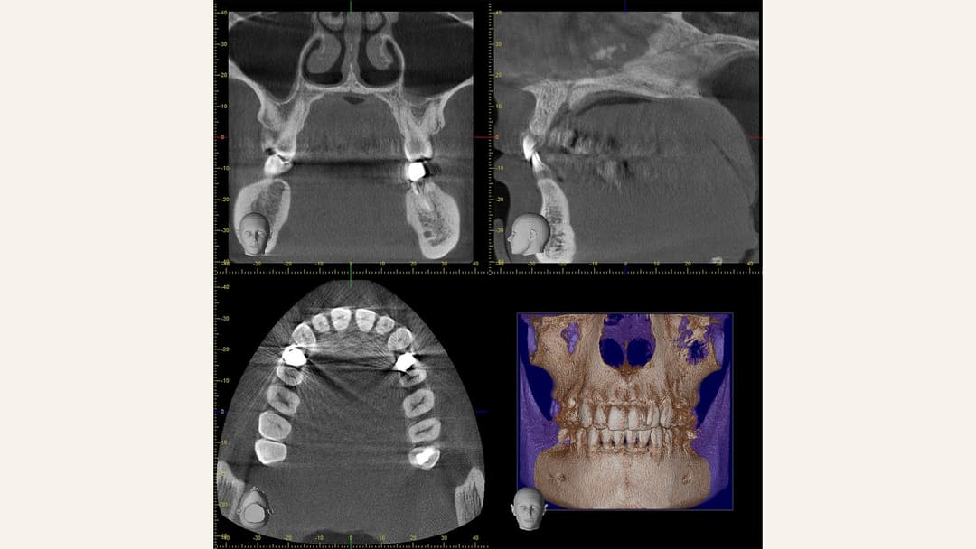
Conventional X-rays are flat images in which structures overlap. CBCT (cone-beam computed tomography) reconstructs the teeth, jawbone and nerve canals in three dimensions, allowing the dentist to examine the anatomy slice by slice.
Clinically, CBCT is commonly used to: measure bone height and width and locate the inferior alveolar nerve before dental implant placement; assess complex wisdom teeth lying close to the nerve; find hidden canals or evaluate root-end lesions in difficult root canal cases; and plan orthognathic surgery in three dimensions.
Radiation dose: what the numbers actually mean
Everyone receives natural background radiation every day (cosmic rays, the environment, and so on) — about 2 to 3 millisieverts (mSv) a year, so a small amount daily. Dental imaging doses can be understood as “how many days of background radiation this is equivalent to”:
・A small dental film (digital): roughly a few hours to one day of background radiation
・A panoramic OPG: roughly one to three days of background radiation
・A cephalometric X-ray: similar to an OPG, or lower
・Dental CBCT (small field of view): roughly several days to two weeks of background radiation, depending on the field setting
・For comparison: a hospital head CT is measured in millisieverts and is substantially higher than a dental CBCT; the cosmic radiation on a single Hong Kong–London flight is already higher than one OPG
Digital sensors also require much less dose than traditional film. Combined with accurate aiming and collimation (limiting the beam to the area needed), doses from modern dental imaging have fallen substantially.
What about lead aprons? International guidance has been updated in recent years: a 2024 recommendation from the American Dental Association (ADA), issued with the American Academy of Oral and Maxillofacial Radiology, takes the view that with modern digital equipment and accurate field limitation, routine use of lead aprons and thyroid collars does little to reduce dose and can even obscure the image so that a retake is needed. Local regulations and guidance do not necessarily move at the same pace, and day-to-day practice follows local requirements. If you have questions about shielding, or a particular circumstance (for example pregnancy), you are welcome to raise it with the dentist before the examination — we will explain the arrangements in place at the time.
The underlying principle does not change: image only when clinically needed, and keep the field as small as practical — that remains the most effective way to manage dose.
Not sure which imaging you need?
Imaging is recommended by the dentist based on your clinical situation. Message us on WhatsApp or call 3956 9387 and we will arrange a suitable assessment appointment.
Digital 3D intraoral scanning: how does it differ from traditional impressions?
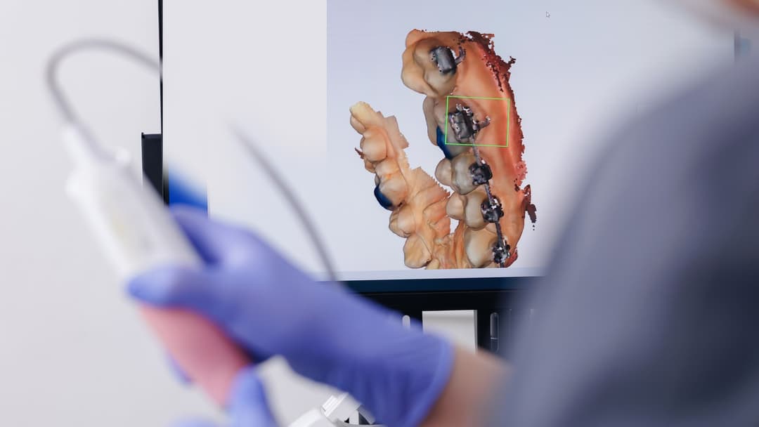
Traditional impressions require impression material to set in the mouth, which some patients find uncomfortable or gag-inducing. Digital 3D intraoral scanning instead moves an optical scanning wand around the mouth, converting the teeth and gums into a three-dimensional digital model in real time.
The digital model feeds directly into the design of crowns, bridges, porcelain veneers and clear aligners, removing several physical-model steps. That said, each method has its place — for some complete denture cases, traditional impressions still perform well — and your dentist will recommend the approach that suits the treatment.
Why choose Home Dental for imaging and digital dentistry?
✓ Panoramic, cephalometric, CBCT and digital 3D intraoral scanning equipment on site — imaging and consultation completed in one place
✓ Images are interpreted by your treating dentist for the treatment in question; cross-specialty cases (implants, orthognathic surgery, complex wisdom teeth) can be assessed jointly
✓ Findings and treatment options are explained in detail before treatment, and fees are clearly set out after assessment
✓ Available at both our Causeway Bay and Tsuen Wan clinics
Book your professional dental care at Home Dental
You can arrange a same-day consultation via:
• The WhatsApp button on this page (recommended) — reach us instantly
• Calling 3956 9387 (during office hours)
WhatsApp EnquiryCall +852 3956 9387
To learn more, you may also read: Dental implants — what to know and consider.