Key takeaways
A root canal file breaking inside the tooth (instrument separation) is a recognised complication in the endodontic literature — most likely in curved, long or calcified canals, which are features of the tooth itself, not necessarily a sign of error. The fragment is sterile metal, typically just a few millimetres long; if the canal was already properly cleaned and disinfected before the fracture, sealing the fragment in with the filling can often achieve a good long-term result — removal is not automatic, and forcing out a deep fragment can do more harm than leaving it. Options include entombment with monitoring, bypassing, microscope-assisted retrieval and apical surgery, recommended after CBCT and microscope assessment. In short: no need to panic — assess first, then decide.
"The dentist says a file broke during my root canal and part of it is still in the tooth." Most patients feel alarm first and anger second. This article is here to help you take a calmer view — because in most cases the situation is less serious than it sounds. Let us explain what instrument separation actually is, why it happens, what it really means for your tooth, and what your options are.
What is instrument separation — and why does it happen?
Root canal files are extremely fine metal instruments that must rotate and flex inside narrow, curved canals. In canals that are particularly curved, long, narrow or calcified (hardened and constricted), metal fatigue rises sharply and a file can fracture, leaving a fragment in the canal — known professionally as instrument separation.
To be clear: instrument separation is a known, documented complication in the endodontic literature, and it is heavily influenced by the tooth's own anatomy — curved roots, long roots, calcified canals are factors no dentist controls, so its occurrence does not by itself mean the treating dentist made an error. The professional standard is to inform the patient promptly, record it, and explain the follow-up options.
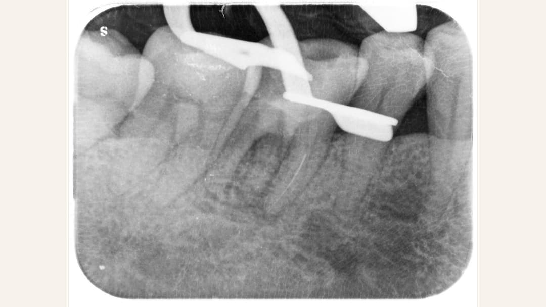
How serious is a retained fragment? Usually less than you imagine
Many patients worry about metal left in the body. The fragment is sterile stainless steel or nickel-titanium, typically only a few millimetres long — it does not "rust and cause infection" and is not itself a source of disease. The only real question is: does the fragment block cleaning and disinfection of the canal portion beyond it?
Timing is everything. If the fracture happened after the canal was already thoroughly cleaned and disinfected, sealing the fragment within the root filling frequently achieves a good long-term outcome — the literature and clinical experience both support this. Conversely, forcing out a fragment lodged deep beyond a curve requires removing more tooth structure, and can weaken the tooth and add trauma. "It must come out" is simply not the correct starting point.
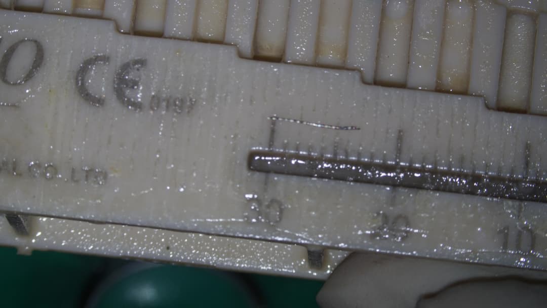
The options — recommended per your case
Entombment with monitoring: if the fracture occurred after the canal was substantially cleaned and there are no infection signs, the most reasonable course is often to complete the filling, seal the fragment in place, and monitor with periodic X-rays. This is not "making do" — it is a legitimate, evidence-supported option.
Bypassing: rather than removing the fragment, a path is negotiated alongside it so the canal beyond can still be cleaned, disinfected and filled, sealing the fragment within the filling.
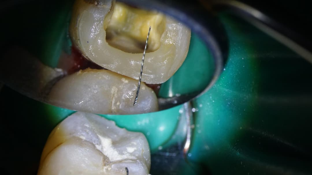
Microscope-assisted retrieval: under the operating microscope, ultrasonic instruments loosen the dentine around the fragment so it can be retrieved. Feasibility is higher when the fragment sits nearer the crown with a straighter canal and good visibility; for fragments deep beyond the apical curve, the difficulty and cost to the tooth rise sharply, and your dentist will weigh with you whether it is worth it.
Apical surgery (apicectomy): when the fragment lies near the root tip with persistent infection and other approaches are not feasible, treating the affected root end surgically can be considered.
There is no one-size-fits-all answer — the dentist weighs fragment position, canal anatomy, infection status and the tooth's restorative value, and explains the trade-offs before treatment. The best approach is to sit down with your endodontist — or a dentist you trust — and talk through each option's feasibility, risks and costs one by one, so you make the decision together, fully informed before anything is done.
The role of CBCT and the microscope
Managing instrument separation is above all about seeing clearly: a CBCT scan establishes the fragment's position, canal curvature and root-end status in three dimensions before treatment — informing whether retrieval, bypassing or monitoring is the sounder choice — while the operating microscope provides high magnification and coaxial lighting for direct vision deep in the canal. For these cases, specialised equipment is the decisive factor: a millimetres-long fragment deep in a canal simply cannot be managed safely by the naked eye. Retrieval is performed with fine ultrasonic tips that gently vibrate the dentine around the fragment loose at high frequency — rather than drilling it away — with one goal: completing the treatment with the least possible loss of tooth structure. These assessments take place within our root canal treatment and endodontic specialty services.
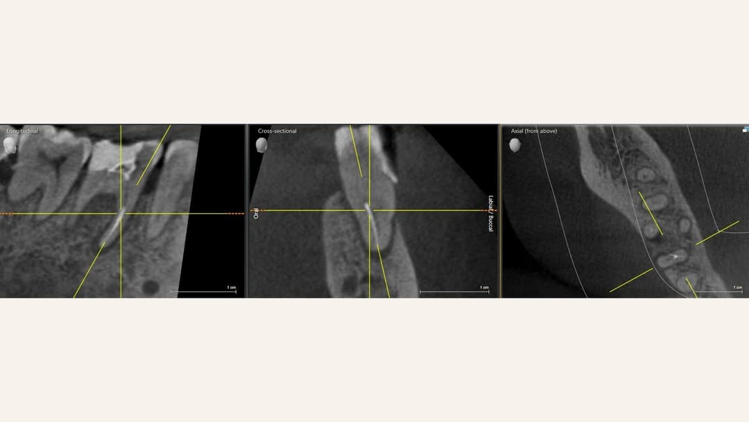
What to bring to an assessment
If the separation happened during treatment at another clinic and you would like a second opinion, bring: recent X-rays (if available), treatment records or a referral letter (if available), and a note of roughly what stage the treatment had reached. The more complete the records, the more accurate the assessment. Requesting these records from your original clinic is entirely normal practice. If the situation involves a treatment dispute, see also dental incident support.
What the outcome looks like
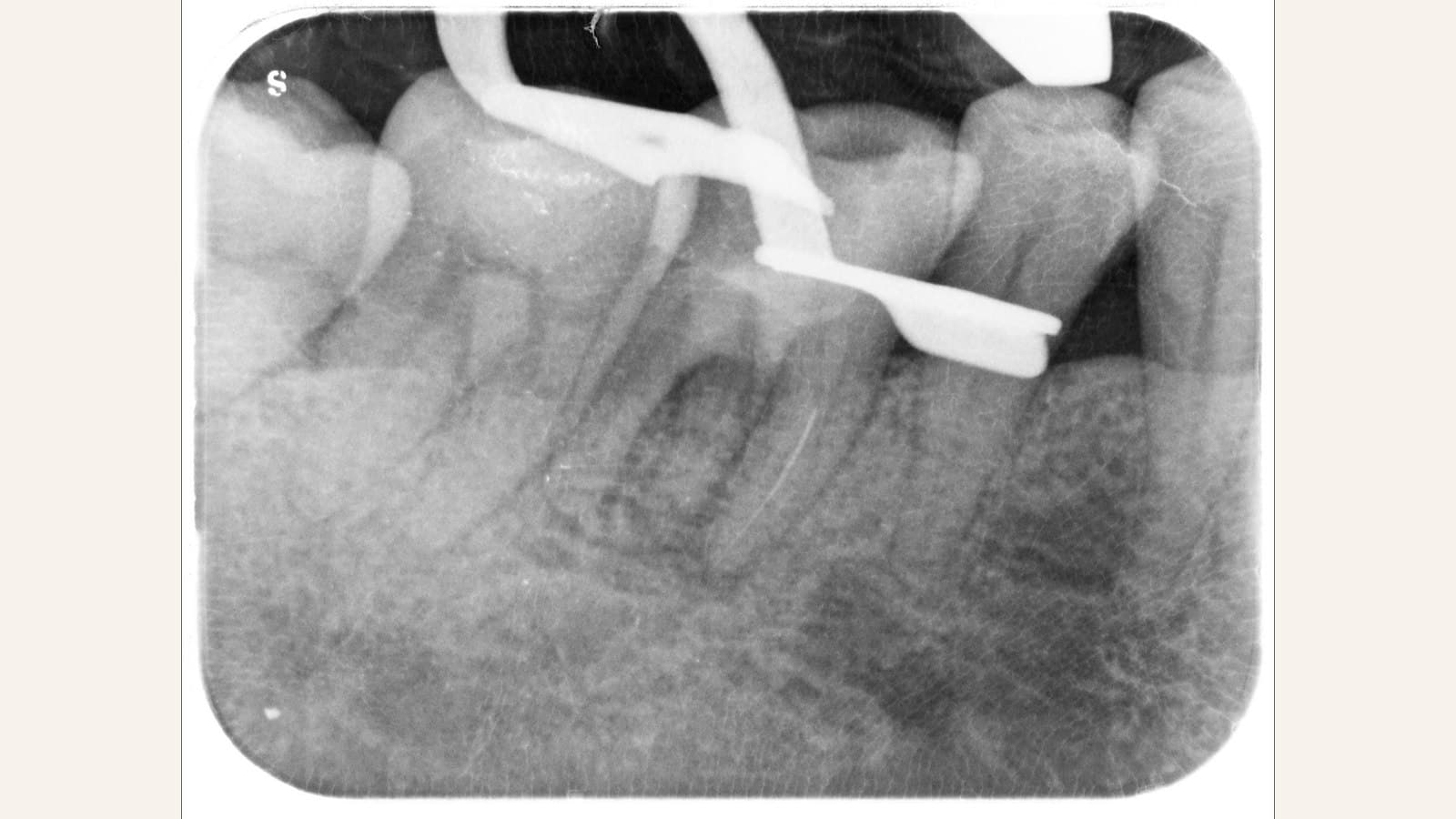
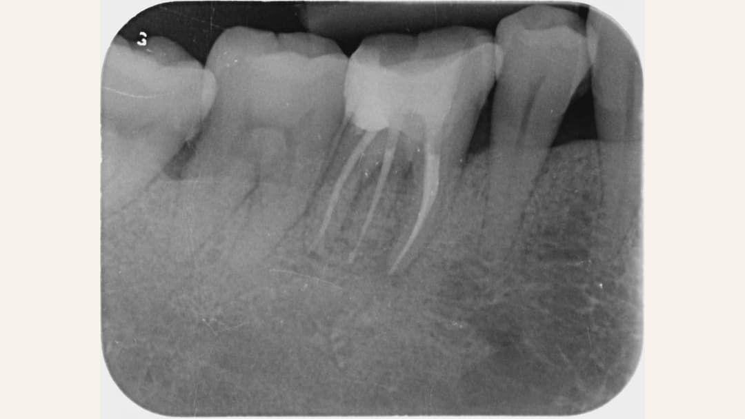
Whether the fragment is retrieved and the canal filled, or entombment is chosen after assessment, the goal is the same: a clean, sealed canal system and a tooth kept in long-term function. Below, a real case after completed root canal filling — dense, even obturation, tooth preserved.
Why choose Home Dental to assess a separated instrument?
✓ Endodontic specialty service equipped with operating microscopes and ultrasonic instruments
✓ On-site CBCT for immediate three-dimensional assessment of fragment position and canal anatomy
✓ Every option — entombment, bypass, retrieval, surgery — explained with its feasibility and trade-offs; we will not recommend retrieval unnecessarily; fees clearly set out after assessment
✓ Assessments available at both Causeway Bay and Tsuen Wan
Book your professional dental care at Home Dental
You can arrange a same-day consultation via:
• The WhatsApp button on this page (recommended) — reach us instantly
• Calling 3956 9387 (during office hours)
To learn more about root canal treatment, read: Root canal therapy.