Key takeaways
A lump, a white or red patch, or an ulcer that hasn't healed after two weeks — most oral lesions turn out benign, but neither the eye nor an X-ray can say so with certainty. The only way to know is a biopsy — and the biopsy itself is not frightening: the wound is usually small, closed immediately with a stitch or two, and most patients feel only mild discomfort with simple medication. The spectrum of oral lesions is wide — harmless fibromas; jaw cysts that quietly displace teeth and erode bone; "benign but aggressive" tumours like ameloblastoma; chronic immune conditions such as lichen planus; potentially malignant patches; and oral cancer. Telling them apart is exactly what pathology does. Every removed tissue is sent for histopathology as standard, and anything that won't heal must be checked: found early, treatment is far more conservative.
"There's a painless lump in my mouth — does it matter?" "This ulcer hasn't healed in three weeks — is it just heat?" These questions come up daily, and the answer cannot be guessed. This article explains the wide spectrum of oral lesions, why a dentist recommends a biopsy, what the procedure actually involves (far less daunting than most imagine) — and why "most are benign" and "it must be checked" are not contradictory statements.
Why biopsy? Aren't the eye and the X-ray enough?
No. Very different conditions can look alike: a white patch may be harmless frictional keratosis, lichen planus, a potentially malignant lesion, or an early cancer; a dark shadow on an X-ray may be a simple root-end cyst or an aggressive tumour. Appearance and imaging give clues; tissue gives the diagnosis. A pathologist examining the cells under the microscope is what turns "it might be" into "it is" — and that pathology report is the foundation of every treatment decision that follows. The biopsy is precisely the step that decides the next step: once the report confirms the lesion's nature, you can be told with confidence whether further treatment is genuinely needed — or whether regular observation and review is in fact the best course. Without it, any decision to treat or not to treat is guesswork.
For the same reason, tissue removed at surgery — cysts, lumps — is always sent for histopathology as standard practice: even when a lesion looks confidently benign in the clinic, laboratory confirmation is what completes the treatment.
The spectrum of oral lesions: from entirely benign to needing early action
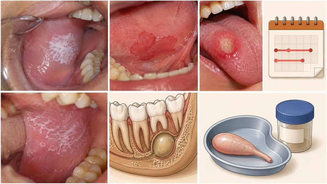
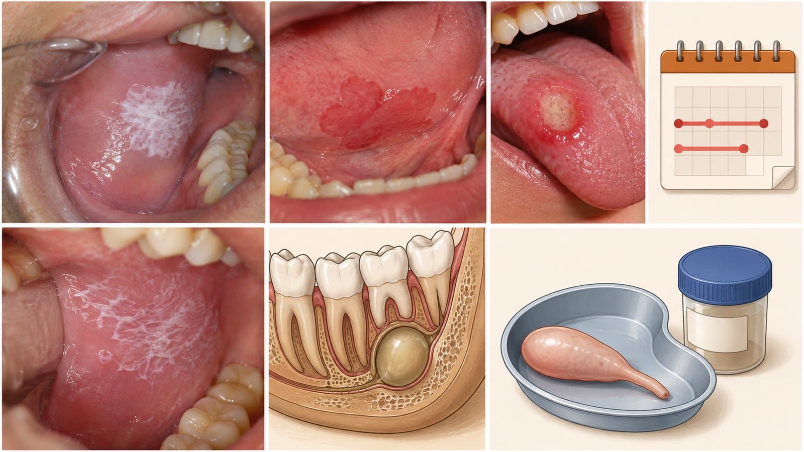
① Common benign lesions: fibromas (firm nodules from chronic cheek-biting or friction), mucoceles (blister-like swellings, typically on the lower lip), ordinary aphthous ulcers — these make up the majority of oral lesions. Treatment is simple, and post-removal histopathology confirms all is well.
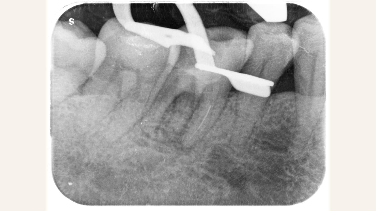
② Odontogenic cysts — benign, but the cost of waiting can be high: such as radicular cysts (linked to a dead tooth nerve, often managed alongside root canal treatment) and dentigerous cysts (commonly around unerupted wisdom teeth). Cysts are usually benign — but they enlarge silently: pushing and tilting the roots of neighbouring teeth and eroding jawbone. Left long enough, the consequences can be loosening and loss of multiple teeth and extensive jawbone destruction — at which point treatment means not just cyst removal but major bone grafting and a long rehabilitation. Caught early, by contrast, the surgery is small and recovery quick — and the removed cyst lining is always sent for histopathology, because certain cyst types need closer follow-up.
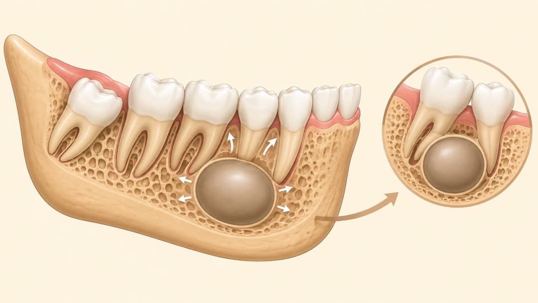
③ Benign but aggressive tumours: the classic example is ameloblastoma — pathologically "benign", yet it grows aggressively, destroys jawbone extensively, and recurs if not removed with an adequate margin. Lesions like this are exactly why "benign" never means "ignore it": diagnosed early by biopsy, treatment can be far more conservative; discovered late, the surgery becomes much larger.
④ Chronic immune-mediated mucosal conditions: the mouth's lining has its own chronic immune diseases — most commonly oral lichen planus: lace-like white striations on the cheek lining, sometimes with sensitivity, burning or erosion; certain autoimmune blistering diseases (such as pemphigoid and pemphigus) can also present in the mouth. These conditions are not cancer, but biopsy is needed to confirm the diagnosis (some require special immunofluorescence testing), so treatment can properly control symptoms — and because individual forms carry a small malignant potential, long-term regular monitoring after diagnosis matters too.
⑤ Potentially malignant lesions: mucosal changes such as leukoplakia (white patch) and erythroplakia (red patch) — some carry malignant potential, with red patches generally higher-risk than white. Such lesions do not necessarily become cancer, but a biopsy is needed to grade the cellular changes, and the report guides whether removal or regular monitoring follows.
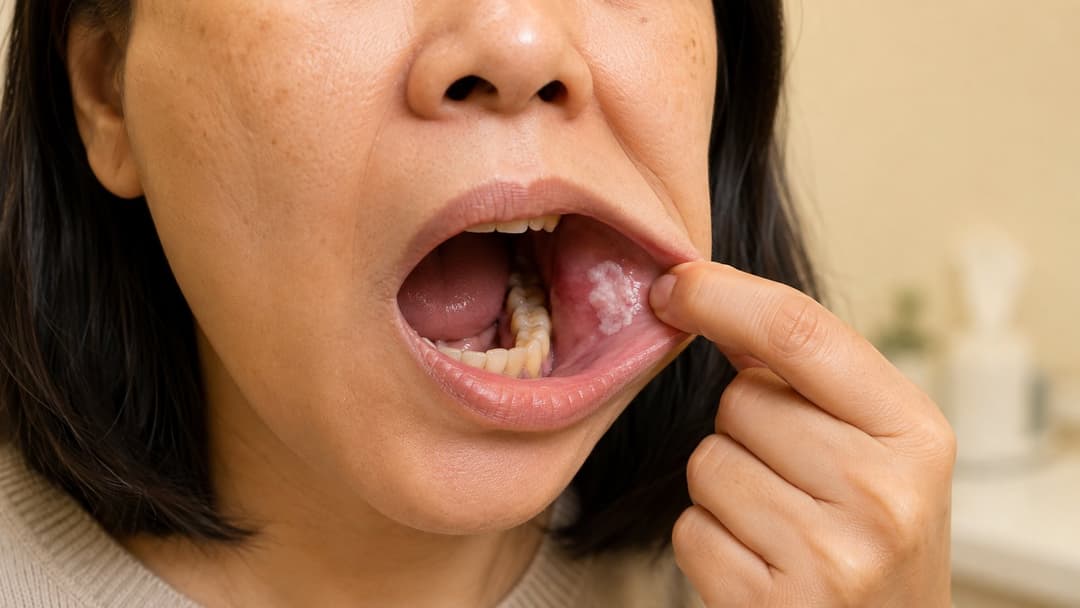
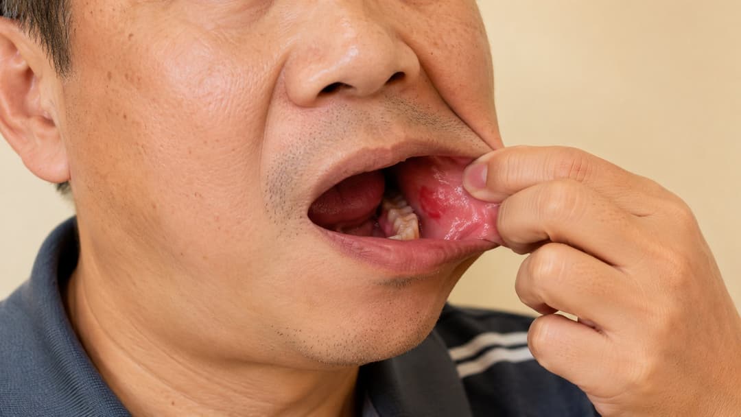
⑥ Oral cancer: malignant tumours of the tongue, floor of mouth, cheek lining, gums and other sites. Risk factors include smoking, alcohol and betel-nut chewing. The difference between early and late detection — in treatment complexity and in outcome — is enormous; and early oral cancer is often painless, which is why vigilance about "not normal, not healing" matters so much.
The two-week rule: when to get checked
Any oral ulcer or lesion that has not healed within two weeks should be examined by a dentist — stop waiting to "see if it settles". Also worth attention: persistent white or red patches, unexplained lumps or thickening, unexplained numbness, teeth loosening without gum disease, and an extraction socket that will not heal. Most of these turn out benign — but "most" is not "certainly", and a check is what buys certainty.
What does a biopsy actually involve? Less than you fear
Far simpler than most people imagine: under local anaesthesia, an oral & maxillofacial surgeon takes a small sample of the lesion (incisional biopsy) or removes a small lesion whole (excisional biopsy) — and in cyst cases, the entire cyst lining removed at surgery is submitted. The wound is usually small and closed immediately (typically a stitch or two); most patients experience only mild discomfort, well managed with simple medication, healing within days with daily life largely unaffected. For deep cysts or anxious patients, the procedure can be done under dental sedation (monitored anaesthesia care) for complete comfort.
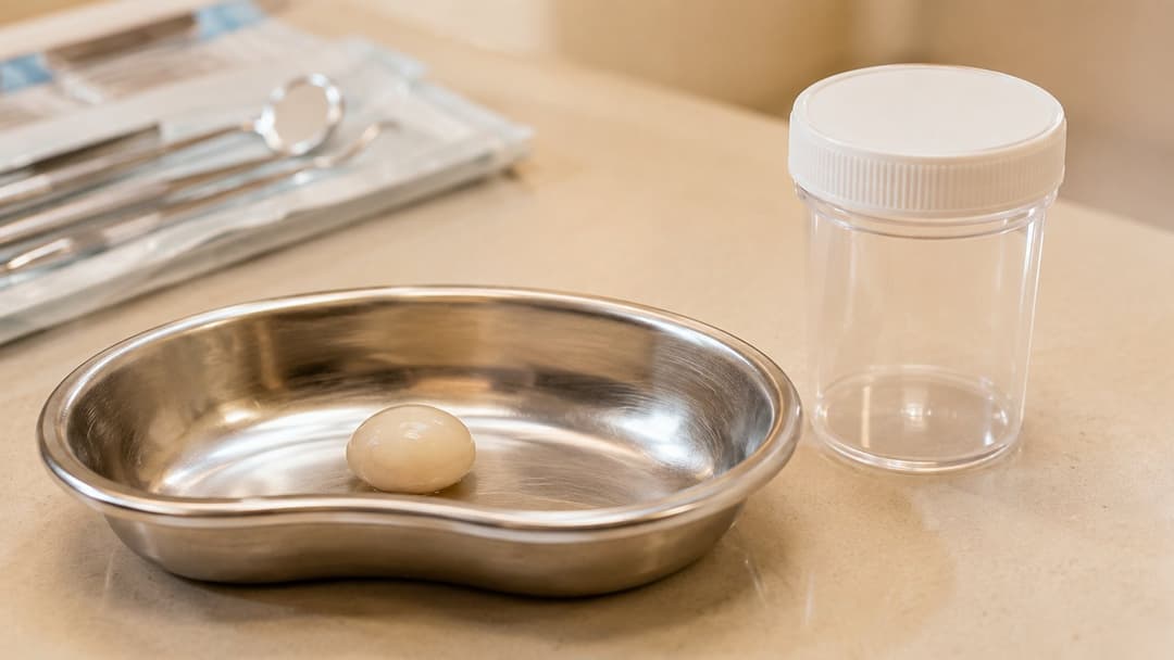
The tissue goes to a pathologist, and the report typically takes one to two weeks. Your dentist then explains the result and the follow-up plan — in most cases the result is benign and follow-up is simple; where the report calls for further treatment, knowing early is precisely the value of having checked. For deeper or larger lesions, imaging (X-ray / CBCT) maps the extent before the biopsy is planned.
Why choose Home Dental for oral lesion assessment?
✓ Biopsies and cyst removal led by well-trained oral & maxillofacial surgeons experienced in managing the full range of these conditions
✓ Treatment planned strictly on the pathology report, always favouring the most conservative, tissue-preserving approach — never over-treatment
✓ Deep cysts and anxious patients can be treated under dental sedation (monitored anaesthesia care) in our licensed Day Procedure Centre
✓ Affiliated arrangements with several private hospitals — higher-risk or extensive cases can be arranged in a hospital setting, ensuring the fullest support for complication management
✓ On-site X-ray and CBCT for immediate assessment; all removed tissue sent for histopathology as standard, with the report explained in detail; fees set out after assessment; Causeway Bay and Tsuen Wan
Book your professional dental care at Home Dental
You can arrange a same-day consultation via:
• The WhatsApp button on this page (recommended) — reach us instantly
• Calling 3956 9387 (during office hours)
Related services: Oral & maxillofacial surgery · Dental sedation.